MERSCOPE Protein Stain Reagent Kits

Enabling True Multiomic Spatial Measurements

Multiomic spatial measurements that combine transcript and protein detection, with analyte location are critical in understanding cell interactions, signaling, and overall tissue behavior. Vizgen’s MERSCOPE Protein Stain Reagent Kits enable true multiomic measurements by co-detecting proteins and transcripts in a single MERFISH experiment.

Figure 1

Protein Co-detection updated figure 1

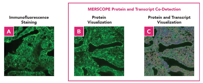
Figure 1: A. Immunofluorescence staining of cytokeratin proteins in fresh frozen human uterus cancer tissue with mouse pan-Cytokeratin antibody. B. C.MERSCOPE co-detection of transcripts from a panel of 244 genes and cytokeratin proteins using MERSOPE protein stain kit and pan-cytokeratin antibody in fresh frozen human uterus cancer tissue. B.  Visualization of detected proteins. C. Visualization of detected transcripts and proteins.

Request a Quote

MERSCOPE Protein Stain Reagent Kits 

High quality co-detection of transcripts and proteins in a single experiment 

Vizgen's Protein Stain Reagents Kits provide all the necessary reagents to co-detect up to 5 proteins with a single MERFISH experiment. 

MERSCOPE Protein Stain Reagent Advantages:

  • MERFISH technology provides multiplexed, error robust transcript detection 
  • Equivalent staining between MERSCOPE protein reagent staining and immunofluorescence staining
  • True co-detection of proteins and transcripts on the same tissue sample in a single MERFISH experiment
  • High correlation with bulk RNA-seq data for high, medium and low expressing genes 
Vizgen Reagent Product Family 1

High Quality Protein Transcript Co-detection Enables Expanded Applications

Accuracy and sensitivity are critical features in generating biologically meaningful spatial genomics data. The MERSCOPE Protein Stain Kits provide superior accuracy and sensitivity to ensure successful, impactful spatial data generation.

Identify Tissue Structures and Characterize Regional Gene Expression

Figure 2

Protein tissue structure

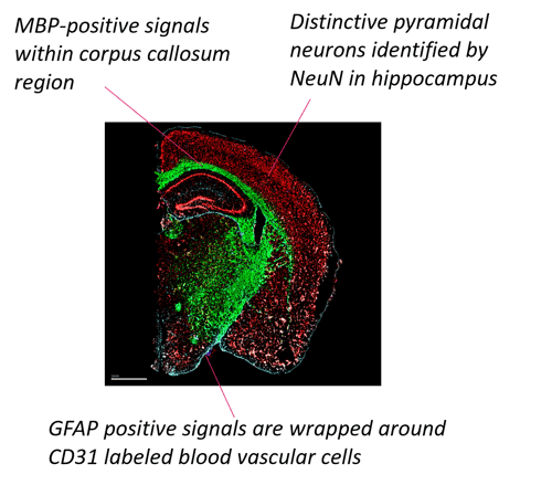
Figure 2: A merged image of MERSCOPE protein co-detection of 468 genes and five protein staining: NeuN-red, Iba1-white, CD31-Blue, MBP-green, and GFAP-light blue in mouse brain. 

Utilize Protein Markers to Classify Cell Types


Figure 3

Protein classify cell types no border

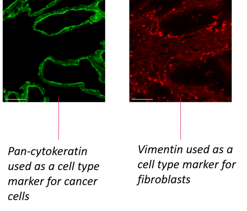
Figure 3: Staining of individual protein in human colon cancer. Pan-cytokeratin used for classification of cancer cells. Vimetin for classification of cell types.

MERSCOPE FFPE Anti-body Co-staining Data Release:
Unraveling Immuno-Oncology Signatures in Human Uterine Cancer with MERSCOPE

 Cancer immunotherapy has emerged as a promising avenue for treating various malignancies, including human uterine cancer. Research to understand the interplay between immune cell populations and tumor microenvironments is critical for developing effective therapies. For this data showcase, we demonstrate the capability of spatial multi-omics profiling with the Vizgen MERSCOPE® Platform. Specifically, we combined protein staining for CD45RA and CD45RO, key immune cell markers involved in T-cell differentiation and activation, with MERFISH to provide a multi-dimensional view of the tumor microenvironment within FFPE human uterine cancer samples.

ProteinCodetection MERSCOPE MasterPiece 1